


Home / Training / Manuals / Atlas of breast cancer early detection / Cases
Atlas of breast cancer early detection
Go back to the list of case studies
.png) Click on the pictures to magnify and display the legends
Click on the pictures to magnify and display the legends
| Case number: | 027 |
| Age: | 57 |
| Clinical presentation: | Postmenopausal woman with average risk of developing breast cancer presented with a slowly growing lump in the left breast for 6 months before presentation. Examination revealed a hard lump (5 × 3 cm) in the retroareolar region of the left breast. |
Mammography:
 |  |
| Breast composition: | ACR category c (the breasts are heterogeneously dense, which may obscure small masses) | Mammography features: |
| ‣ Location of the lesion: | Left breast, central portion of the breast, central zone, anterior third |
| ‣ Mass: | |
| • Number: | 0 |
| • Size: | None |
| • Shape: | None |
| • Margins: | None |
| • Density: | None |
| ‣ Calcifications: | |
| • Typically benign: | None |
| • Suspicious: | None |
| • Distribution: | None |
| ‣ Architectural distortion: | Present |
| ‣ Asymmetry: | Focal |
| ‣ Intramammary node: | None |
| ‣ Skin lesion: | None |
| ‣ Solitary dilated duct: | None |
| ‣ Associated features: | Architectural distortion |
Ultrasound:
 |  |
| Ultrasound features: Left breast, central portion of the breast | |
| ‣ Mass | |
| • Location: | Left breast, central portion of the breast |
| • Number: | 1 |
| • Size: | 5.4 × 3.1 cm |
| • Shape: | Irregular |
| • Orientation: | Parallel |
| • Margins: | Circumscribed |
| • Echo pattern: | Hypoechoic |
| • Posterior features: | No posterior features |
| ‣ Calcifications: | None |
| ‣ Associated features: | Minimal internal vascularity |
| ‣ Special cases: | None |
BI-RADS:
BI-RADS Category: 4B (moderate suspicion of malignancy)Further assessment:
Further assessment advised: Referral for cytologyCytology:
 |
| Cytology features: | |
| ‣ Type of sample: | FNAC |
| ‣ Site of biopsy: | |
| • Laterality: | Left |
| • Quadrant: | Central |
| • Localization technique: | Palpation |
| • Nature of aspirate: | Whitish |
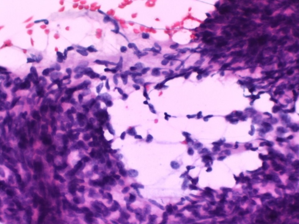
| ‣ Cytological description: | Smear shows large sheets of tightly cohesive benign ductal epithelial cells. There is no significant nuclear pleomorphism. Many large stromal fragments are also seen with both plump and slender spindle cells. A few bare nuclei are also seen |
| ‣ Reporting category: | Benign |
| ‣ Diagnosis: | Benign phyllodes, but frozen section advised to check margins of surgical excision |
| ‣ Comments: | None |
Histopathology:
Simple mastectomy
 |  |
| Histopathology features: | |
| ‣ Specimen type: | Simple mastectomy |
| ‣ Laterality: | Left |
| ‣ Macroscopy: |
|
| ‣ Histological type: | On histology, sections from the tumour show features of a benign phyllodes tumour (i.e. stromal hypercellularity and benign glandular elements seen in the tumour). No microscopic features of malignancy such as increased mitotic activity, marked atypia of the stromal cells, and infiltrative margin, necrosis, or haemorrhage are seen. Sections from the two firm areas seen grossly show only dense sclerosed stromal tissue. There is no evidence of epithelial hyperplasia in sections from the remaining breast |
| ‣ Histological grade: | |
| ‣ Mitosis: | |
| ‣ Maximum invasive tumour size: | |
| ‣ Lymph node status: | |
| ‣ Peritumoural lymphovascular invasion: | |
| ‣ DCIS/EIC: | |
| ‣ Margins: | Specimen 1: The posterior margin of the tumour is very close to the skeletal muscle fibres but separated from them by fascia and fibroadipose tissue. All the surgical margins of the resection are free of tumour. The skin is uninvolved.
Specimen 2 labelled as deeper margin revised: Sections show only skeletal muscle tissue. There is no evidence of phyllodes tumour in these sections. Specimen 3 labelled as revised base: Sections show skeletal muscle fibres with fibroadipose tissue. There is no evidence of phyllodes in these sections |
| ‣ Pathological stage: | |
| ‣ Biomarkers: | |
| ‣ Comments: |
Case summary:
| Postmenopausal woman presented with left breast lump. Diagnosed as area of focal asymmetry and architectural distortion of suspicious morphology in left breast, BI-RADS 4B on imaging and as benign phyllodes tumour on cytology and histopathology. |
Learning points:
|